Joseph Toynbee (1815-1866) of England wanted
to do more work with otology. He dissected
more than 2000 temporal bones and formed the
collection which became known as the Toynbee
Collection in the Museum of the Royal College of
Surgeons. In 1860, his work "Disease of the
Ear" was published. It contained
information on the dissection of diseased
ears. Toynbee showed that stricture of the
Eustachian tube was not a common affliction
since he had only one out of his 1523
dissections. He noted that the Eustachian
tube was not permanently open, but lightly
closed, and that it became opened only during
such movements as swallowing or
yawning. In one of his dissections,
Toynbee recognized a fistula of the external
semicircular canal and he pointed out that
infection could extend to the brain by way of
the labyrinth. Tonybee was one of the first
to describe otosclerosis (a condition
characterized by chronic progressive deafness)
and he recognized it in 160 cases.
The Eustachian tube (pharyngotympanic tube)
connects the middle ear cavity with the
nasopharynx. It aerates the middle ear system
and clears mucus from the middle ear into the
nasopharynx. Opening and closing functions of
the eustachian tube are physiologically and
pathologically important. Normal opening of the
eustachian tube equalizes atmospheric pressure
in the middle ear; closing of the eustachian
tube protects the middle ear from unwanted
pressure fluctuations and loud sounds.
Mucociliary clearance drains mucus away from the
middle ear into the nasopharynx, thus preventing
infection from ascending to the middle ear.
The eustachian tube in the adult is
approximately 40 mm long and is directed
downward, forward, and medially from the middle
ear. It consists of 2 portions, a lateral third
(13 mm), which is a bony portion arising from
the anterior wall of the tympanic cavity, and a
medial two thirds (27 mm), which is a
fibrocartilaginous portion entering the
nasopharynx. The tube opens about 1.25 cm behind
and slightly below the posterior end of the
inferior turbinate.
The eustachian tube in infants measures
approximately 18 mm in length. It is about half
the size of the adult eustachian tube and is
generally more horizontal and less angulated.
The bony portion is relatively longer and wider
in diameter, the nasopharyngeal end of the
cartilaginous portion lies more inferiorly.
The only active muscle that opens the
eustachian tube is the tensor veli palatini,
which promotes ventilation of the middle ear.
The eustachian tube also functions to protect
the middle ear from excessive sound pressure,
and nasopharyngeal secretions. The eustachian
tube helps drain the middle ear during opening
and closing by pumping secretions from the
middle ear; clearance of secretions also occurs.
An understanding of the anatomy and physiology
of the system can aid the clinician in
understanding the role of eustachian tube
dysfunction in the cause and pathogenesis of
middle ear disease and the possible contribution
of allergy to this disease.
Abnormal or impaired eustachian tube
functions (ie, impaired opening or closing,
defective mucociliary clearance) may cause
pathological changes in the middle ear. This in
turn can lead to hearing loss and other
complications of otitis media. These
pathological changes include recurrent acute
otitis media and otitis media with effusion.
Chronic retraction of the tympanic membrane may
also lead to middle ear atelectasis and
subsequent adhesive otitis media. A retraction
pocket of the tympanic membrane secondary to
chronic eustachian tube dysfunction may
eventually evolve into cholesteatoma and
potentially serious complications.
Bartolomeo
Eustachi publie le premier, en 1707, une
définition décrivant avec
exactitude la trompe d'Eustache:
"La portion de la trompe dont
l'extrémité semblable à un
coin vient au contact de l'os temporal est
manifestement cartilagineuse et
extrêmement épaisse; en
réalité la structure de la portion
opposée n'est pas exactement
cartilagineuse mais elle a je ne sais quoi de
membraneux et se termine en s'amincissant.
Cependant à l'extrêmité
interne de ce méat, situé en
regard de la cavité nasale, le cartilage
est solide, augmenté de volume, et
recouvert par la tunique muqueuse nasale et on
le voit planté comme un portier au bord
du méat. Sa forme n'est pas ronde, mais
un peu affaissée et forme deux angles
...."
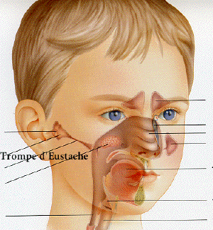
La trompe d'Eustache est un conduit
fibrocartilagineux et osseux qui assure la
communication entre le rhino-pharynx ou cavum et
l'oreille moyenne. Il mesure environ 4
centimètres. Son orientation
générale est oblique chez l'adulte
et presque horizontale chez l'enfant. Le fait
que la trompe d'Eustache, la caisse tympanique
et l'antre soient quasiment dans le même
plan, favorise chez l'enfant l'apparition
d'otites séromuqueuses (les
sécrétions rhino-pharyngées
remontent vers la trompe principalement en
position couchée).
L'ouverture de la trompe d'Eustacbe est
commandée par un ensemble de muscles qui
relient le vélo-pharynx et la trompe
d'Eustache. Le vélo-pharynx est
formé du voile du palais et
d'éléments musculaires constituant
les parois postérieures et
latérales du pharynx. Celles-ci sont
formées de fibres musculaires qui
présentent une double disposition (des
fibres longitudinales à action
élévatrice et des fibres
circulaires à action constrictive).
Le voile du palais prolonge en bas et en
arrière la voute palatine. C'est une
cloison musculo-membraneuse mobile et
contractile. Il sépare l'oro-pharynx du
cavum et intervient dans la déglutition,
la phonation et le bâilllement.
Les muscles péritubaires
interviennent dans le mouvement de la trompe
d'Eustache. Les plus importants sont: le muscle
péristaphylin externe (muscle primordial
dans l'ouverture tubaire, il joue aussi un
rôle important dans la déglutition)
et le muscle péristaphylin interne. Le
péristaphylin interne met la trompe
d'Eustache en position d'ouverture et le
péristaphylin externe l'ouvre. La
contraction synergique de ces deux muscles est
indispensable.
La trompe d'Eustache a un fonction
d'aération de l'oreille moyenne ou
fonction équipressive. Elle apporte
à l'oreille moyenne l'air venant du
rhino-pharynx, et maintient sa pression
égale à la pression
atmosphérique ambiante.
Physiologiquement, le trompe d'Eustache s'ouvre
à chaque déglutition; sa
non-ouverture aggrave la dépression
ét favorise une la
sécrétion inflammatoire de la
caisse conduisant à l'obstruction de la
trompe d'Eustache. Elle a également une
fonction de drainage de l'oreille moyenne vers
le nasopharynx. Lors de son ouverture, elle
limite la transmission des bruits physiologiques
liés à la déglutition, par
exemple.
Les dysfonctionnements tubaires
réalisent une
dysperméabilité tubaire cause et
conséquence principale de l'otite
séromuqueuse.
Les causes extrinsèques principales
sont l'hypertrophie des
végétations adénoïdes
ou toute cause d'obstruction nasale, les tumeurs
du cavum, les insuffisances
vélo-pharyngées, les troubles de
l'articulé dentaire. Ceux-ci favorisent
une hypertonie permanente des muscles
ptérygoïdiens externes perturbant de
l'ouverture de la trompe d'Eustache lors de la
déglutition.
Anatomiste de talent, Bartolomeo
Eustachi a fait progresser cette science
dans la seconde moitié du XVIe
siècle. Outre la fameuse trompe
d'Eustache, il a révélé
l'existence de la valvule qui porte son nom, des
surrénales, du canal thoracique.
Le grand oeuvre de Bartolomeo Eustachi
devait être un traité d'anatomie
« De dissensionibus ac controvesiis
anatomicis ». Il devait comporter 47
planches anatomiques, dessinées avec
l'aide de Pier Matteo Pini, richement
détaillées et
légendées. Seulement 8 planches
furent publiées de son vivant. Les 39
autres, perdues, ont été longtemps
recherchées. Elles ont été
retrouvées 162 ans plus tard chez un
descendant de Pier Matteo Pini. Publiées
en 1714 sous le titre « Tabulae anatomicae
Bartolomaei Eustachi quas a tenebris tandem
vindicatas » (illustrations anatomiques de
Bartolomeo Eustachi sauvées de
l'obscurité), elles font de leur auteur,
avecVésale, l'un des pères de
l'anatomie moderne.