Case Report: We describe a
59-year-old gentleman with abnormal involuntary
movement of paralyzed right upper limb during
yawning 2 weeks following ischemic stroke of
left middle cerebral artery territory.
Discussion: This is a rare
post-stroke phenomenon and its
pathophysiological mechanism is poorly
understood but this entity highlights possible
preserved extrapyramidal pathway which might
help in rehabilitating stroke survivors.
Introduction
The rare phenomenon of abnormal involuntary
movement of paralyzed upper limb in association
with yawning following stroke was termed as
parakinesia brachialis oscitans (PBO) by
Walusinski et al. [1]. Only few cases
have been described in the literature and there
are many unanswered question of this clinical
condition. We report a patient of PBO after
acute infarction in left middle cerebral artery
(MCA) territory.
Go to: Case Report A 59-year-old
right-handed gentleman presented in the
Neurology outpatient department (OPD) with
history of acute onset right hemiparesis and
motor aphasia of 4-weeks duration. He was a
known diabetic, hypertensive and smoked 1 packet
of cigarettes per day for 30 years. There was no
past or family history of stroke or myocardial
infarction. His CT scan of brain did not reveal
any intra-parenchymal haemorrhage or any
evidence of infarct. He was treated by his
physician soon after the stroke and was
gradually improving.
About 2 weeks following the stroke, his
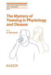
relatives noticed an involuntary lifting of the
hemiplegic right arm with yawning. On observing
the patient at OPD, this abnormal movement was
confirmed. He was still having weakness of his
right sided limbs (Video 1). The movement was
synchronous with every episode of yawning. The
movement consisted of slow progressive abduction
of shoulder, flexion and mild supination at
elbow, associated with occasional dorsiflexion
of wrist. The phenomenon was observed with each
episode of yawning (Video 2). It was of same
duration as the episode of yawning. It started
shortly after the start of yawn with the hand
getting lifted up during the entire inhalation
phase of the yawn and then quickly returned to
its original position during the brief
exhalation phase. The patient remained fully
conscious throughout the movement. There were no
other associated movements noticed in his face,
left upper limb or lower limbs.
Besides motor aphasia there was spasticity
of his right upper and lower limb and normal
tone in his left side. There was right sided
upper motor neuron facial weakness and right
hemiparesis with power of grade 2/5 in upper
limb and 3/5 in lower limb (Medical Research
Council) and normal power on left side. There
were exaggerated tendon reflexes and positive
Babinski sign on right side and equivocal
plantar response on the left.
MRI brain showed a chronic infarction in
left lateral frontal cortex and white matter
extending to operculum, precentral gyrus,
sylvian cortex and basal ganglia with evidence
of haemorrhagic transformation (Figures 1 and
_and22). MR angiography revealed loss of flow
signal in intracranial left internal carotid
artery and left MCA (Figure 3). A1 segment of
left anterior cerebral artery was hypoplastic
with patent anterior communicating artery.
Doppler ultrasound of bilateral carotid and
vertebral arteries showed atherosclerotic
plaques in bilateral carotid arteries without
significant occlusion. Electrocardiogram and
echocardiogram were suggestive of left
ventricular hypertrophy without any evidence of
atrial fibrillation, intracavitary thrombus or
any valvular pathology.
He was managed conservatively with
antiplatelets, high dose statins,
antihypertensives, oral hypoglycaemic agents,
physiotherapy and speech therapy. Since then the
patient is on regular follow up in Neurology
OPD. There has been significant improvement of
his motor power and speech functions in the last
one year. The abnormal movements are still
persisting, however there has been a decrease in
the degree of lifting of the paretic limb.
Discussion
Cases of movement of paralysed arm in
hemiplegic limbs which were completely
disobedient to the will was known for a long.
However, Walusinski et al. in 2010 proposed the
term parakinesia brachialis oscitans (PBO) for
this rare phenomenon [1]. The term
parakinesia, as defined by the authors, means
"an abnormal involuntary movement that acts as a
parasite, caricature or replacement of a normal
movement" and oscitans means "yawn" in Latin
[1].
This movement is typically seen in the
paralysed upper limb in synchrony with the
yawning episode. However, two cases were
reported to have some associated movements in
the hemiplegic lower limb [2] and may be
considered as variants of PBO. These
involuntary, patterned movements usually include
abduction and internal rotation of shoulder,
flexion of elbow and extension of fingers. These
are usually slowly progressive movements which
start with the onset of yawn and continues
throughout the inhalation phase of the yawn
followed by a swift return of the affected limb
to its original position.
In one of the reported cases [3],
there was tremor associated with the movement.
In another reported case, patient could perform
complex purposeful act during movement and also
could wilfully supress it [4]. It
usually takes about 5 seconds for the entire
movement. In most of the cases, these are
observed soon after the acute insult (generally
within first week), however late onset during
the spastic phase may also be seen. There was no
suggestion of any age or gender predilection but
most reported cases of PBO were men. Also, there
was no preference for any laterality or
dominance. These movements tend to disappear
with recovery of motor function in the affected
limb, usually within six months. However, it may
persist for longer periods as seen in our
patient. The most common etiologies which were
found in reported cases of PBO were stroke.
Although both ischemic and haemorrhagic strokes
were found to be associated with PBO, most of
the cases available in literature are ischemic
stroke. Meenakshisundaram et al. (2010) studied
75 patients with abnormal movement during
yawning following acute stroke and reported a
mean onset time of 36 to 38 hours in male and
females, respectively [5]. Associated
movements in hemiplegic limbs during yawning
were minimal and observed in 78.6%, which was
significantly more in males (83% vs 70%), in
those with hypotonia (87% vs 61%)), and in
proximal joints (72% vs 29%) irrespective of
limb. It was also more common in left-sided (94%
vs 64%) hemiplegics, and in the upper limbs (91%
vs 83%). Table 1 shows cases of PBO reported by
r The two common sites of lesion were internal
capsule and pontomedullary region. Other sites
include centrum semiovale, frontal subcortex and
total MCA territory infarct.
Yawning is a physiological phenomenon seen
in almost all vertebrates. The paraventricular
nucleus of hypothalamus plays the key role in
yawning through its connections with hippocampus
and brain stem areas such as reticular formation
and locus coeruleus [3]. The final
executive pathway of yawning involves motor
nuclei of cranial nerves V, VII, IX, XI and XII
and C1&endash;C4. Neocortical brain areas
possibly exert an inhibitory effect on these
subcortical structures.
The pathophysiology of PBO is still not
clear. Walusinski et al. proposed that in PBO
there is interruption of corticospinal,
corticobulbar and corticopontocerebellar
pathways [1]. However, the
proprioceptive loop carrying signals between the
paleocerebellum, lateral reticular nucleus and
motor anterior spinal horn remains functional.
So during yawning, strong contractions of
respiratory muscles lead to generation of
proprioceptive signals that reaches the lateral
reticular nucleus. Motor signals from lateral
reticular nucleus then travel via extrapyramidal
pathways to the anterior horn cells of
C4&endash;C8 causing movement of the affected
upper limb.
Interruption of cortico-pontocerebellar
pathway is essential for PBO so that
proprioceptive loop remains disinhibited. This
interruption can occur at two levels, at
internal capsule affecting the first order
neurons or at the level of pons affecting the
second order neurons, the two most common sites
of lesion for PBO.
Other suggested pathophysiologic mechanism
for PBO include disinhibition of subcortical
structures by cortical damage that may release
the reticular brainstem formation interconnected
with motor pathways and may be activated by
stimuli such as yawning [6]. Other
explanation includes that an "emotional motor
system" may be responsible for the movement of
paralysed upper limb and yawning would activate
it as a consequence of emotional state related
to drowsiness [7].
Our patient developed PBO two weeks after a
large haemorrhagic infarct in the left lateral
frontal cortex and white matter extending to
operculum, precentral gyrus, sylvian cortex and
basal ganglia interrupting the descending
pyramidal tracts. The preserved proprioceptive
loop in presence of interrupted corticospinal,
corticobulbar and corticopontocerebellar
pathways seems to be the most likely explanation
for PBO in our patient. Although there was a
gliosis suggesting old infarct in the opposite
hemisphere, it is difficult to associate that
with the phenomenon.
Conclusion
PBO is a rare and interesting phenomenon
seen commonly in post-stroke patients. The exact
pathophysiological mechanism is still unknown
but it generates tremendous curiosity.
Understanding this complex phenomenon might help
in rehabilitation of stroke survivors by
manipulating preserved extrapyramidal
pathways.