Yawning has been rarely described in
association with seizures and not previously
documented by video-EEG. We present a
48-year-old woman with a long history of
non-dominant for speech hemisphere seizures and
post-ictal yawning. Yawning was irresistible,
forceful and often repetitive. We reviewed the
few similar epileptic cases described in the
literature and discussed the possible
mechanisms.
Yawning is a common physiological event in
mammals. It may increase vigilance when
drowsiness occurs and is probably important for
social communication. It has been reported in
different pathological conditions (Daquin et al.
2001). The occurrence of yawning in relation to
epileptic seizures has been rarely described
(Penfield and Jasper 1954, Goldie and Green
1961, Donat and Wright 1991, Muchnik et al.
2003). We report a patient with non-dominant for
speech hemisphere seizures and irresistible,
repetitive and forceful post-ictal yawning.
Case report
A 48-year-old, left-handed woman with no
neurological antecedents began having
intractable epileptic attacks at age 18. She
described five types of seizures:
- Seizure type 1, isolated myoclonic jerks
of the left arm,
- Seizure type 2, left arm numbness,
- Seizure type 3, type 2 progressing to
dystonic posturing of the left arm with
non-forceful head deviation to the left, and
brief unresponsiveness, occasionally followed by
a transitory left arm weakness,
- Seizure type 4, sudden falls without
warning,
- Seizure type 5, rare secondarily
generalized tonic-clonic seizures (GTCS).
Surface EEG recordings showed interictal
epileptic activity over the right
centro-parieto-temporal regions. Her
neurological examination was normal. Medical
history was remarkable for asymptomatic
hepatitis C. At 30, she underwent invasive EEG
studies, which disclosed epileptiform
abnormalities over the right parietal operculum.
Brain CT and MR imaging were normal.
Neuropsychological evaluation revealed right
hemisphere dysfunction (sodium amobarbital test
confirmed a left hemispheric dominance for
speech). A right inferior parietal and posterior
temporal resection did not lead to improvement.
Pathological studies showed no specific changes.
At 31, she had a second resection at the
temporal edge of the previous operation, again
with no improvement.
In 2004, she underwent further telemetry
recordings. Thirty-eight seizures were recorded:
9 were myoclonic jerks; 6, left arm sensory
seizures (duration, < 10 s); 22, sensory
attacks followed by left hand dystonia, head
deviation and loss of awareness (mean duration,
15 s). No falls were recorded but she had one
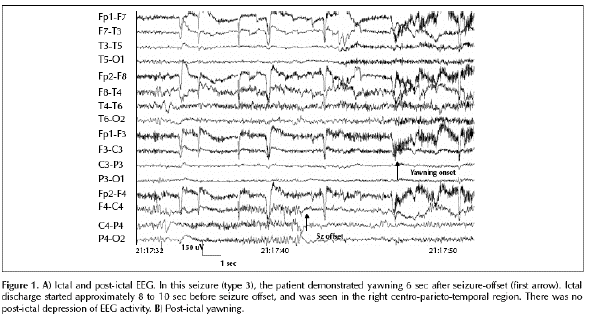
GTCS. Ictal scalp EEC showed no changes with
types 1 and 2, and disclosed low voltage fast
activity over the right centro-parieto-temporal
areas without depression of background activity
with all type 3 seizures (figure 1). Attacks
occurred at random during the day when she was
alert and active, and alpha activity was seen
before and after each of the 37 seizures that
did not generalize. Complete blood count,
electrolytes, renal and thyroid function tests
were normal, but hepatic enzymes were mildly
elevated. Oxygen blood saturation and CO2 were
not measured during yawning, but routine venous
blood parameters were normal.
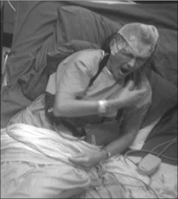
Yawning.
In 19 type 3, and in two type 2 seizures,
she had repetitive, irresistible and forceful
yawning starting from one to 30 seconds (mean,
8.5 s) after the seizure offset. No yawning was
detected with myoclonic jerks, and the single
GTCS was not included in the analysis. Yawning
lasted from five to 60 seconds. There was no
change of cardiac rhythm. There was no obvious
change in blood pressure, respiration and no
skin changes occurred during these episodes.
Yawning started with a deep inspiration through
her wide-open mouth (figure 2). She was alert
during all yawning episodes. She received
oxcarbazepine 1,800 mg/d during telemetry and
her valproate was gradually withdrawn. Her
husband had witnessed similar paroxysmal yawning
for approximately six years occurring typically
at the end or just after type 3 seizures when
she received different medications singly or in
combinations. Physiological yawning was observed
when the patient was tired or bored, with a
quality similar to her peri-ictal yawning except
for lack of clustering and irresistibility. We
found no episodes of peri-ictal yawning when
reviewing the earlier (1986 and 1987) prolonged
surface and intracranial video-EEC studies. Her
seizures remained the same over the years,
including those followed by yawning. Repeated
high quality MR failed to show a structural
lesion, except for postsurgical changes.
Discussion
We describe a woman with repetitive yawning
as an unusual, isolated, autonomic, post-ictal
phenomenon. Yawning appeared immediately or
seconds after cessation of seizures, was
irresistible and was observed after most (86%)
of the focal sensory-motor and some (33%)
sensory attacks. We cannot explain why yawning
appeared late (approximately 24 years after
onset of her seizures and 11 years after her
second surgery) during the course of her
disorder.
Physiology of yawning. Yawning is a
complex spatiotemporal reflex, and neural
structures involved are presumably located in
the brainstem near respiratory and vasomotor
centers, and those that control facial mimicry,
mastication, throat movements and stretching.
The purpose and mechanism of yawning are not
well understood. Three theories have been
proposed: a relation to respiration, alertness
or communication (Daquin et al. 2001). The
hypothalamic paraventricular nucleus contains
nerve endings that belong to the
incerto-hypothalamic dopaminergic system and
project to oxytocinergic neurons located in this
nucleus. These in turn project to the
hippocampus, pons and medulla oblongata and
mediate the expression of yawning. Oxytocinergic
neurons can be modulated by several
neurotransmitters and neuropeptides, such as
dopamine, oxytocin, prolactin, excitatory amino
acids, acetyicholine and nitric oxide to name
the most important ones (Argiolas and Melis
1998). Oxytocin injected in the CAl field of the
hippocampus induced yawning (Melis et al. 1986).
Acetylcholine and cholinomimetic drugs can
induce yawning in rats and muscarinic
antagonists can prevent it. Finally, prolactin
facilitates and opioids inhibit yawning.
Yawning and epilepsy.
We found only six, well documented cases in
the literature where yawning was reported in
association with epilepsy (table 1). Penfield
and Jasper (1954) described two adult patients
with autonomic seizures and yawning. The first
had a tumor infiltrating the left temporal lobe
and ipsilateral basal ganglia. She had seizures
beginning with a headache associated with
yawning, hiccup, urge to void and focal sensory
phenomena. In the second patient, attacks were
ushered in by sweating of her hands and scalp
and a prickling sensation in the scalp and back,
and with repetitive yawning, palpitations,
visual distortions and weakness. No EEG was
reported. Golgie and Green (1961) described an
adolescent with "petit mal" and found an
association between yawning and 3 per second
spike and wave discharges. Donat and Wright
(1991) reviewed unusual variants of infantile
spasms in 11 children (13% of their patients
with infantile spasms), and described one, with
an unspecified brain malformation, who had a
variant consisting of yawning with generalized
decremental activity on EEC. This yawning
variant and the child's more typical infantile
spasms responded to ACTH. Finally, Muchnik and
colleagues (2003) described two patients who had
temporal lobe epilepsy and yawning. In the
first, yawning occurred during drowsiness and
preceded focal seizures. The patient had
independent, bilateral, temporal interictal
epileptiform discharges, but imaging was not
reported. The other, a 17-year-old woman with an
insulin-dependent diabetes, had a normal MRI,
complex partial seizures and secondary
generalization with yawning during the postictal
period. EEC also showed bilateral temporal
spikes.
Three mechanisms might explain peri-ictal
yawning. First, activation of the brainstem
structures related to a change in the level of
alertness. In our patient, yawning was never
caused by a change in her level of alertness.
This mechanism however, could be a factor in the
two patients of Muchnik and colleagues (2003).
Second, a direct activation of brainstem
structures by the epileptic discharge. The fact
that yawning appeared immediately or just a few
seconds after a right centro-parieto-temporal
epileptic discharge and seizure offset, suggests
a fast operating process that activated the
brainstem structures. We can not explain why
yawning did not occur during the attack. Third,
a seizure-mediated secretion of endogenous
neurohumoral substances such as prolactin or
oxytociri may cause yawning. The latter is less
likely since a prolactin surge should only occur
after a few minutes (Pritchard et ai. 1983), and
activation of oxytocin in the hypothalamic
paraventricular nucleus was found to occur only
1.5 hours after kainicinduced seizures in rats
(Sun et al. 1996). We did not however, measure
plasma concentration of prolactin or oxytocin in
our patient. There is no evidence that her
medications were responsible for yawning since
the patient's husband and she herself did not
notice any change in the yawning pattern over
the previous six years when taking various
drugs.
In keeping with previous reports implying
preferential non-dominant hemisphere for the
induction of autonomic peri-ictal symptoms such
as spitting, water drinking, vomiting, urinary
urge or coughing (Baumgartner et ai. 2001),
yawning in our patient occurred after seizures
arising in her non-dominant hemisphere. However,
such nondominant lateralization may not be
absolute, since a patient with yawning and
seizures related to a left temporal tumor was
reported (Penfield and jasper 1954). It is
remarkable that forceful yawning has been so
rarely described in association with seizures.
D
Figure 1 A) Ictal and post-ictal EEG.
In this seizure (type 3), the patient
demonstrated yawning 6 sec after seizure-offset
(first arrow). Ictal discharge started
approximately 8 to 10 sec before seizure offset,
and was seen in the right
centro-parieto-temporal region. There was no
post-ictal depression of EEG activity. B)
Post-ictal yawning.
References
Argiolas
A, Melis MR. The neuropharmacology of
yawning. Euri Pharmacol 1998; 343:1-16.
Baumgartner C, Lurger S, Leutmezer F.
Autonomic symptoms during epileptic seizures.
Epileptic Disord 2001; 3: 103-16.
Penfield
W, Jasper H. In: Epilepsy and the functional
anatomy of the Human Brain. Boston, Mass:
Little, Brown & Co, 1954:416-8.
Pritchard 3rd PB, Wannamaker BR, Sagel J,
Nair R, DeVillier C. Endocrine function
following complex partial seizures. Ann NeuroI
1983; 14: 27-32.
Sun Q, Pretel S, Applegate CD, et al.
Oxytocin and vasopressin mRNA expression in rat
hypothalamus following kainic acidinduced
seizures. Neuroscience 1996; 71: 543-54.
Specchio N,
Carotenuto A et al. Ictal yawning in a
patient with drug-resistant focal epilepsy:
Video/EEG documentation and review of literature
reports.Epilepsy Behav
2011