- As late as twenty years ago physiologists
and clinicians agreed in declaring the cortex of
the brain to be functionally, homogeneous.
Flourens's experiments had decisively negatived
Gall's very ingenious but purely hypothetical
conception, and any effort to prove localization
would, at that day, have seemed like a reversion
to a system already tried and condemned. It was
freely admitted that, from experiments made on
pigeons, one might infer the mode of brain
functionment in man. Medicine was under the yoke
of the then dominant teachings of physiology,
nor was there so much as a thought of reaction.
Clinical observers, indeed, had long before
known that motor troubles consequent on a lesion
of the brain imply localization of such lesion
in the hemisphere on the side opposite to that
paralyzed; but that was then the sum of the
topographical diagnosis.
-
- Broca, in 1863,
showed that the impairment of the power of
articulate speech, which he calls aphemia, is
connected with a brain affection that is always
localized in a clearly circumscribed region of
the left hemisphere. At first the fact was
called in question. When proofs had been
multiplied in its favor men contented them
selves with simply admitting it, little noting
that this very definite localization was a first
attack on Flourens's doctrine, which must now
undergo revision. But the topographical anatomy
of the cerebral convolutions was then too little
known to enable one to "find his bearings" on
the surface of the brain, and the reaction
against Flourens's ideas would at that time have
met with insurmountable obstacles.
-
- The thorough researches of Leuret and
Gratiolet, and of
their successors, Ecker, Broca, Gromier, by
making us acquainted with the morphology of the
external surface of the brain, removed these
first anatomical difficulties. The experiments
of Fritsch and Hitzig in Germany, in 1870, and
shortly afterward those of Ferrier, in England,
modified the ideas which prevailed. They showed,
on the one hand, that the gray matter of the
brain is not incapable of excitation, as had
been supposed; that electric excitation of this
gray matter calls forth motor reactions; on the
other hand, they prove -an important point- that
the effects produced differ according to the
part of the cortex that is excited. From that
date, properly speaking, began researches into
motor localizations in the brain. Since then
such researches have been prosecuted in two
directions; for while the physiologists
reproduced, with various results, the
experiments of Fritsch, of Hitzig, and of
Ferrier, the clinicians were also at work. And I
may be permitted to say that the researches in
this latter direction began in France, and that
I have had some share in them. My first
researches, made jointly with Professor Pitres,
then my interne, were the starting-point for
studies that have been for ten years prosecuted
with remarkable activity in France, where a
great number of investigators have contributed
their share of facts, in England by Jackson and
Ferrier, in Germany by Nothnagel.
-
 - On considering how far we have advanced in
the study of localization in the cortex while
puriuing these two pathsexperimentation on
animals and anatomo-clinical observation of
man-one is struck with the fact that while among
clinicians there is perfect agreement, at least
on the essential points, amorg the physiologists
there is marked disagreement. The divergence of
views is due, perhaps, mainly to the fact that
the experimenters have cared less about
determining the relations between a given
affection and a lesion of one or another part of
the cortex, than about discovering the inner
mechanism of the relation between the two. That
which, in the eyes of the clinician, whose
thoughts are ever of diagnostics, is the point
of capital importance, thus becomes an accessory
datum for the experimenter, who thinks more
about theory. Now, the theories that have been
advanced, one after another, to account for the
phenomena observed to follow excitation or
destruction of the cortex are as numerous as
they are uncertain. Take the fundamental facts
alleged by Fritsch, Hitzig, and later by
Ferrier, viz., that excitation of certain parts
of the gray matter determines localized
convulsions; that, on the contrary, ablation of
these parts produces paralysis; these facts,
while admitted in their general tenor, have been
interpreted in very different ways. According to
some writers, Ferrier, for instance, the cortex
comprises true motor centers; others, as Hitzig,
Fritsch, Schiif, Munk, hold the excitable points
to be sensitive centers, excitation of which
determines movement in virtue of a sort of
reflex action, while destruction of these
centers produces paralysis through loss of
conscious, sensibility. Many phvsiologists, as
Tamburini, Luciani, and Seppeli, hold this
"excitable zone "to be both motor and sensitive.
Vulpian held that it is simply the place of
convergence or influences emanating from all the
other parts of the encephalon, and that it has
no activity of its own. Finally, according to
Dr. Brown-Squard, the excitable points of the
cortex have neither motor nor
sensitivo-sensorial functions; excitation
applied to them does but traverse them, passing
on to organs of movement situate lower down;
their destruction does not act by suppression,
but by irritation at a distance. Such is the
theory of dynamogenic, or inhibitory, action at
a distance. As has been justly remarked by
François Franck:
-
- "It must be admitted that all the
interpretations now conceivable are absolutely
provisional ; nay, it were rash and illogical to
believe that any question whatever touching the
mechanism of the brain, and in particular this
one, has been definitely settled."
-
- Certainly the study of these questions is by
no means void of interest, and the clinician may
not stand indifferent toward the efforts made to
determine the instrumental process whereby a
given lesion of the cortex produces such or such
a convulsion, such or such a paralysis. But he
must not forget that this determination is a
secondary task; and, in any case, theoretic
considerations cannot fairly be suffered to call
in question the positive teachings of
anatoino-clinical observation.
-
- Then, it is to be borne in mind that
experimentation with animals that are nearest to
man, still more with those far removed from man
on the zoological scale, cannot, however
faultless its technique, however definite its
results, solve finally the problems raised by
the pathology of the human brain. In brain it
is, above all, that we differ from animals. That
organ attains in man a degree of development and
of perfection not reached in any other species.
Its functions become complex, while at the same
time its morphology undergoes important
modifications. Now, it is perfectly clear that
as regards questions of localization
morphological details are of the first
importance. As for functions, even if we take
account only of those common to men and animals,
they are not performed in all in the same way.
The higher an organism stands in the animal
scale, the more strictly are the purely reflex
functions subordinated to the functions of the
higher centers. A decapitated frog performs with
its legs co-ordinated automatic movements ; not
so a decapitated dog. In the dog, brain lesions,
even of considerable extent, produce only
incomplete paralysis, often passing away, while
in man the like lesions cause incurable
functional troubles. These examples are enough
to show that, particularly as regards brain
functions, the utmost reserve is necessary in
drawing inferences from animals to man. The
results of experimentation, however ingenious,
however skillfully conducted, can give only
presumptions more or less strong, but never
absolute demonstration.
-
- Hence, the only really decisive data
touching the cerebral pathology of man are, in
my opinion, those developed according to the
principles of the anatomo-clinical method. That
method consists in ever confronting the
functional disorders observed during life with
the lesions discovered and carefully located
after death. This is the method that enabled
Laennec to throw light on the difficult subject
of diagnosing pulmonary affections, and it has
also materially helped the diagnosis of diseases
of the liver, kidneys, and spinal cord. To it, I
may justly say, do we owe whatever definite
knowledge we have of brain pathology. As for the
localization of certain cerebral functions, here
this method is not only the best, but the only
one that can be employed. What light, for
instance, could experimentation have thrown upon
the question as to the seat of the functions of
speech-functions which are special to man?
-
- No doubt observations restricted to the
domain of man, and deprived of the powerful
lever of experimentation, may, at first sight,
seem doomed to play a subordinate and
inconspicuous role, but that is so only in
appearance. As I had occasion to write, some
twelve years ago:
-
- "The conditions of a truly spontaneous
experiment in man are presented every day in
pathological circumstances. To profit by them,
we have only to learn to comply with the
necessities of a situation no doubt very
different in many respects from that which
experiment purposely brings about in the animal,
but which is not always more complex. If it is
true that observations made, in the light of
physiology, on man in disease, usually require
more time, more patience, than corresponding
studies of animals under experiment ; if it is
true that in man the conditions of the phenomena
cannot be, as they are in the laboratory, either
modified or reproduced at the will of the
observer ; so, too, is it true tnat disease
often determines in the body of the patient
lesions more strictly limited to one organ or
one tissue; in other words, more systematic and
more compatible with persistence of life, and
with the integrity of functions not directly
concerned ; consequently they lend themselves
better to methodical and protracted analysis
than do mutilations produced in animals by even
the most skillful physiologist." (Revue
Scientifique, Nov. 11, 1876).
-
- But in order to be employed with profit,
anatomo.clinical observations must not be
gathered at hap-hazard. On the contrary, they
have to be tested methodically and classified
according to certain rules that I have taken
pains to define from the beginning of my studies
on cerebral localizations. It is plain, for
instance, as I have elsewhere said, that
irritative lesions are a very different thing
from destructive lesions; nor must we confound
lesions newly produced (accompanied, as they
almost necessarily are, by phenomena having
their seat either near by or at a distance) with
old lesions, in which the morbid process being,
in a measure, at an end, is now clinically
represented only by the mere inactivity of the
parts that have been diseased or destroyed. Just
because these distinctions have not been
sufficiently noted by authors, most of the old
observations are useless as regards the question
of localizations. When we add that in these
observations the designation of the lesioned
convolutions is commonly vague and lacking in
precision, it is seen that such data give but
little light. Hence, as Nothnagel justly says of
the many cases of brain lesions that are
recorded, having been collected in the course of
ages, unfortunately only a very few can bear
criticism or warrant conclusions. But while we
must distrust the old data, we may well accept
those which in these latter years have been
carefully collected by authors who understand
the exigencies of the anatomo-clinical method.
By taking their stand upon these clinicians have
been able to formulate the propositions to which
I am now to call attention, and which form the
groundwork of topographical diagnosis in the
pathology of the brain. In this summary
statement I intend absolutely to avoid reference
to facts that are not perfectly established, for
instance, those bearing on sensitive
localizations; I will mention only such as may
be regarded as firmly and deft nitely
settled.
-
- When a brain lesion, whether cortical or of
any other sort, is accompanied by motor
paralysis, the seat of the paralysis is always
on the side opposite to that of the lesion. This
proposition is universally accepted by
physicians, and in clinics it may be said to
have the force of a law. I would not have
referred to this elementary, truth had not some
physiologists in these latter days ventured to
call it in question, or at least sought to
lessen its diagnostic value by citing in
opposition to it alleged contradictory facts.
But when these observations are subjected to
criticism, it is easily seen that they have no
such force as they have been credited with. In
the record of a clinical case there may easily
occur an error as to the side affected "right"
instead of "left" and vice versa. To some such
lapsus, as I can show, is to be referred the
apparent anomalousness of some, at least, of the
facts alleged in opposition to the law of
chiasm; hence, in my opinion, no weight is to be
attached to cases, even modern cases, in which
authors have not taken pains to insist
explicitly on this anomaly.
-
- And even were it proved that in a few cases,
that are surely exceptional, the paralyss and
the lesion producing it are both on the same
side of the body, it would be necessary, before
drawing an inference from such facts, to make
sure that they are not to be explained by an
abnormal arrangement of the nerve conductors.
This calls for a few words of explanation. We
know that the centrifugal, or motor, fibers
proceeding from the brain decussate, those of
the right crossing those of the left side at a
certain point in their course before they enter,
first, the spinal cord and then the muscles.
This decussation takes place at the level of the
Pyramids of the bulb it gives the reason why a
lesion of the right side of the brain produces
paralysis of the left side of the body, and vice
versa. But normally time decussation is
incomplete for though most of the motor fibers
that constitute the pyramid pass into the spinal
cord of the opposite side, some of them take the
straight course and enter the anterior spinal
cord of the same side. These fibers are, under
ordinary conditions, very few in number. But it
may happen, in case of an exception anatomic
arrangement, that the fibers taking the straight
course are more numerous than those which cross.
Of course in such a case a lesion of the brain
would be explained by an anomal of structure,
but that would give no ground of inference
against the law of decussation, which still
holds good in the immense majority of cases.
Even granting, therefore -a thing that has yet
to he proved- that this law is subject to
exceptions, these exceptions are so rare that,
as far as clinical diagnosis is concerned, we
may leave them out of account, and hold it for a
well-established truth that a paralysis of
cerebral origin presupposes a lesion of the
hemisphere of the opposite side. If I have
mentioned incidentally the objections brought
against a proposition long since become classic
in nerve pathology, it was in order to show the
danger of accepting theories, for so a man may
be led to question the most indisputable
clinical facts.
- Turn we now to the study of disorders
consequent on lesions of the cortex. Hemiplegia,
i. e., paralysis of the movements concerned with
the face and with the two members of one side of
the body, is often the consequence of these
lesions. But not all lesions of the cortex are
accompanied by hemiplegia; they are so only when
certain conditions as to the extent of the
lesion, and particularly as to its seat, are
present.
- Now, anatomo-clinical research shows that
even considerable alterations in the gray matter
of the brain cause no motor disturbance when
they are localized in certain regions. These
regions include the sphenoidal, occipital, and
inferior parietal lobes of the pli courbe and of
the insula, the orbital lobule, and the anterior
portion of the first, second, and third frontal
convolutions. These portions of the brain may be
destroyed by softening, may be compressed or
irritated by tumors, by bony splinters, or by
effusion of blood, without in the least
affecting the motility. The case is totally
different if the region destroyed is that
corresponding to the two ascending frontal and
parietal convolutions and the adjoining replis,
viz., the paracentral lobule, the foot of the
first three frontal convolutions, and of the
superior and inferior parietal lobules. In such
cases we always find hemiplegia of the side
opposite to that of the lesion. Here, then, we
have a striking contrast between the gravity of
the symptoms produced by lesions of this zone
and the marked harmlessness, at least the
latency of effects as regards the phenomena of
movement, in the case of lesions to other
portions of the cortex.
- This contrast has been so often noted and
verified in clinics that we can have no
hesitation in admitting the existence, now well
established, of a motor zone in the cortex. This
zone occupies, as we have seen, pretty nearly
the middle portion of the external surface of
each hemisphere; the region anterior or
posterior to this does not, directly at least,
control movements.
-
- This fact, resulting from a careful
comparison of the symptoms observed during life
and of the necroscopic lesions of the cortex, is
further confirmed by anatomo-clinical
observations of another order. The fact is well
known that a nerve fiber degenerates when
separated from its trophic center, which, in the
case of motor fibers, is the motor cell whence
these fibers emanate. On the other hand, we know
that, as a sequel of certain cerebral lesions,
there is developed in the peduncles, bulb, and
spinal cord a degenerescence of the centrifugal
or motor nerve tubes. Turck first brought this
to light in 1851. Soon afterward I verified the
exactitude of this observation in my researches
with Vulpian. The labors of my pupils, Bouchard,
Pitres, Brissaud, in France, and those of
Flechsig, in Germany, have settled the
determining conditions and the topography of
this degenerescence "secondary" degenerescence,
as it is called. Now, not all lesions of the
cortex are equally capable of producing
secondary degenerescence. This special point I
distinctly called attention to in one of my
lectures in 1873. I attach the more importance
to what I said then, because the question of
cortical localizations in man had not yet been
raised, and there could be no suspicion that my
statement was put forward to strengthen a
theory. I said:
-
- "Cerebral lesions en foyer, considered with
respect to the seat they occupy, are not all
equally capable of determining the production of
consequent scleroses. Thus, among these lesions
there are some which are never followed by
descending sclerosis, while others are dead
certain, so to speak, to produce it.
-
- It results from my observations that
extensive superficial softening, when it
occupies either the occipital lobe, or the
posterior portions of the temporal lobe, or the
sphenoidal lobe, or, finally, the anterior
regions of the frontal lobe, is not followed by
consecutive fasciculated sclerosis; while such
sclerosis, on the contrary, regularly appears
when the foyer compromises the two ascending
convolutions (ascending parietal and ascending
frontal) and the contiguous parts of the
parietal and frontal lobes."
- Research has, during the past ten years,
confirmed the exactitude of the foregoing
propositions. We may, therefore, hold it as
certain that secondary degenerescence is never
seen except after cortical lesions; that when
these lesions are in the zone which we have
called the motor zone, that fact of itself
suffices to prove that there is no direct
relation between the motor conductors and the
regions of the gray matter of the brain which we
have called the latent zone, destruction of
which does not cause paralytic effects.
- I might cite more arguments to prove the
reality of the motor zone of the cortex; in
particular, I might reall the fact, demonstrated
by Betz, Mierzezewski, and other authors, that
its structure differs perceptibly from that of
the adjoining regions, and that this zone has a
mode of development peculiar to itself, as shown
by Parrot. But whatever the force of these new
proofs, I do not dwell upon them here, wishing
to stand on the ground of clinical observation
exclusively. On that ground the reality and the
independence of a motor zone are universally
recognized and accepted to-day.
- The question now arises whether this zone is
functionally homogeneous, or whether, on the
contrary, it is not resolvable into distinct
centers, each concerned with the movements of
some special part of the body. Let us see what
is to be learned on this point by the
anatomo-clinical method. Motor paralyses
resulting from lesions of the cortex do not
always assume the form of hemiplegia. They may,
affect the face, the arm, or the leg; in that
case there is "monoplegia", or, as Nothnagel
terms it, "parcellary, paralysis." We must
observe that monoplegia does not necessarily
depend on lesion of the cortex. Besides cases of
monoplegia due to hysteria there are some that
are due to affections of the motor conductors at
points in their course more or less distant from
the convolutions. But we, of course, have to do
only with monoplegia caused by lesion of the
cortex. Now can we, from the localization of a
monoplegia, infer the seat of the affection
which produces it? In 1883 I was led to
conclude, from researches made in conjunction
with Mr. Pitres, that the cortical motor centers
for the two members of the opposite side are
situate in the paracentral lobule and in the
superior two-thirds of the ascending
convolutions; that the centers for the movements
of the lower part of the face are situate in the
upper third of the ascending convolutions, near
the fissure of Sylvius; that very likely the
center for the isolated movements of the arm
lies in the middle third of the ascending
parietal convolution of the opposite side.
Nothnagel reached these same conclusions through
a close analysis of a multitude of facts, and
they are confirmed by observations published
since 1883. This is specially true as regards
the motor center of the inferior members, the
localization of which has been determined with
the utmost exactitude. Sundry recent facts,
particularly those, at my instance, collected by
one of my pupils, Mr. G. Ballet, have, in fact,
shown that the paracentral lobule, with the
uppermost part of the frontal and ascending
parietal convolutions, has specially to do with
the motility of the femur and crus. Hence, when
a case occurs of monoplegia of the inferior
member referable to a lesion of the cortex, we
can affirm that a lesion localized at the points
mentioned is the cause.
-
- Paralysis is not the only manifestation
which enables us to diagnose a lesion of the
cortex and to point out its seat. Alongside of
the "deficit" symptoms, so called, must be
ranged the "excitation" symptoms, which are also
of the very highest diagnostic value in nervous
clinics. The symptoms of this second group are
manifold, and have diverse clinical
significations. I will refer here only to
convulsions of cortical origin, commonly known
as partial epilepsy, or Jackson's epilepsy. A
French author, Bravais, first described, in
1827, under the name of hemiplegic epilepsy, a
variety of epileptiform convulsions that begin
in one member, or on one side of the face, and
which continue to be limited to one of the
lateral halves of the body. Bravais did good
service in isolating the clinical type, but to
Hughlings Jackson, of the London Hospital,
belongs the credit of having shown its
significance and of having brought to light the
relations between partial epilepsy and lesions
of the cortex of the brain. I give a few
details. Partial epilepsy consists sometimes of
simple tremor, again of violent convulsions like
those of true epilepsy, and producing a
condition that may in a moment end in death. The
general characteristic of the spasms is, that
they begin in some isolated group of muscles,
and are thence gradually propagated to other
muscles of the same member, or of the whole
body, before the patient loses consciousness.
The loss of consciousness, however, is not
fatal, as in true epilepsy; it may continue
during the lifetime. Clinicians are now fully
agreed as to the semeiological value of partial
epilepsy, and the latest ohsetvers have
confirmed the fundamental propositions put forth
by me in 1883, in a work in which I had as
collaborateur Mr. Pitres. The following points
may be regarded as fully established: In the
great majority of cases partial epilepsy results
from lesions of the cortex. It but seldom
follows lesions of the central partions of the
brain. The affections which most readily produce
it are limited affections with quick and
progressive evolution (neoplasm, superficial
encephalitis, meningitis, whether acute or
chronic). Partial epilepsy is never observed in
cases of extensive lesions that suddenly
overspread the whole area of the motor zone. The
lesions which produce it are usually in the
motor zone itself, but they may lie outside of
it, provided the affection is capable of
irritating the elements of the motor
convolutions. Thus, then, the topography of the
lesions in this case is less fixed than in the
case of permanent paralysis. That is why
cortical paralysis can exist either with or
without epileptiform convulsions, and vice
versa. The principles that should guide the
clinician are as follows: When, in the intervals
between attacks, the patient subject to
epileptiform convulsions presents no sort of
paralytic phenomena, then the lesion is in the
vicinity of the motor zone of the cortex.
Partial epilepsy begins either in the arm or in
the leg or in the face; but we cannot fix by, an
absolute rule the seat of the cerebral lesion in
its relation to the way the convulsions make
their appearance. Still, the epileptiform
convulsions which begin in the muscles of the
members are generally produced by lesions
situate at the level of the upper two-thirds of
the motor zone, or in its vicinity; those which
begin in the muscles of the face are commonly
the result of lesions occupying the inferior
extremity of the motor zone, or the neighboring
parts.
-
- It is seen that, from the point of view of
exact topographic diagnosis, the epileptiform
convulsions have less vaine than the paralysis,
yet they authorize us to affirm almost with
certainty that they have to do with a lesion of
the cortex.
-
- The first fact clearly established in
cortical localization was, as I have said, that
published by Broca in 1861. That author showed
that disturbance of the faculty of articulate
speech, since called aphemia, motor aphasia, and
logoplegia, depends on a lesion of the foot of
the third left frontal convolution. Latterly,
the question of affections of speech, of
aphasia, has been thoroughly investigated again.
A more searching and a more exact clinical
analysis has shown that there is ground for
thinking that there are four sorts of affections
corresponding to the loss, partial or total, of
one of the four processes by means of which we
enter into relations with our fellow men. These
four processes are speaking, writing, hearing
(of words), and reading. The former two serve us
in expressing and transmitting our thoughts; the
other two serve us in understanding and
receiving the thoughts of others. Each of these
four mental operations may be impaired, either
separately or in conjunction with the others.
Abolition of articulate speech is called Broca's
aphasia, or motor aphasia; abolition of the
power of writing is agraphia; of that of hearing
words is word deafness; of that of reading, word
blindness. Now, as each of these operations has
its physical independence, so each has its
organ, its special center in the cortex. The
lesion which produces motor aphasia is not that
which produces word blindness; the one on which
depends word deafness is not that which causes
agraphia. As yet the precise seat of the four
centers cannot be fixed. As regards two of them
localization may be regarded as certain; for the
other two it is still hypothetical, or, at
least, only probable.
-
- Before we point out these different
localizations it is important to remind the
reader that the left hemisphere of the brain, to
the exclusion of the right hemisphere, governs
the functions of speech. This fact, glimpsed by
Dax, brought clearly to view by Broca wit
respect to aphemia, holds good also with regard
to the other forms of aphasia. Sometimes,
indeed, motor aphasia has been found to result
from lesion of the right hemisphere, but in such
cases the patients are invariably left-handed
persons, that is to say, persons in whom the
right cerebral hemisphere predominates. But such
cases are exceptional; apart from them the rule
is, that we speak, write, read, understand words
with the left brain. Nor is this surprising,
when we consider that, as Gratiolet has shown,
the left brain develops earlier than the right;
hence, when the infant begins to understand and
to utter words, it must use rather the
hemisphere that is better fitted for performing
these functions.
-
- I come now to the localization of the
centers. Two of them, as I have said, those the
destruction of which is followed by agraphia and
word blindness, have not yet been determined
with absolute certainty. The observations
hitherto made must he multiplied, but as far as
they go they lend the highest probability to the
inference that the center which presides over
writing is situate at the foot of the second
frontal convolution, and that the center which
presides over reading occupies the inferior
parietal lobule, with or without the
co-operation of the lobule of the pli courbe. We
have far more decisive data with regard to the
seats of the other two centers. Broca's
researches have proved indisputably that the
center for articulate speech occupies the foot
of the third frontal convolution; the
observations that are brought forward to
contradict this cannot stand criticism. As for
the region of the cortex, lesion of which
produces word deafness, that certainly, as
Nothnagel held as early as 1879, occupies the
first frontal convolution. An analytical
comparison of the seventeen cases recorded by
Seppeli justifies this conclusion.
-
- Such are the most important and the
best-grounded of the localizations discovered
through the anatomo-clinical method. At first
they were not received without calling forth
some opposition; and though most clinicians were
quick to accept these localizations, at least
with regard to motility and the functions of
language, there were, as a matter of course, a
few who rejected them. But the apparently
contradictory facts brought forward by these few
opponents could not bear methodical and rigorous
criticism. To-day one need but consult the
principal medical journals, and in particular
the publications of the Paris Anatomical
Society, in order to form a just estimate of the
number and the force of the data on which are
based the localizations of which I have spoken.
New observations are daily confirming these
localizations, and these observations would
surely be more numerous still, but just now the
publication of facts confirmatory of the
propositions we bave formulated is neglected.
These propositions no longer meet with any
serious contradiction among clinicians. A few
physiologists still call them in question, but
they do so on the ground of certain purely
theoretical conceptions which, as I have shown,
have nothing to do with the very definite
results of the anatomo-clinical method. As
Vulpian justly said:
-
- "All the progress pathology has made remains
as a permanent acquisition, whatever opinion be
held as to the cortical centers of cerebration.
Whether these centers exist or do not exist, it
is henceforth indisputable that a lesion of the
posterior portion of the left third frontal
convolution causes impairment of language; that
a destructive lesion of the superior portion of
the ascending convolutions produces paralysis of
the leg of the opposite side; and that lesion of
the middle parts of the same convolutions is
followed by paralysis of the arm of the opposite
side. No less indisputable is it that certain
irritative lesions of these same parts give rise
to convulsive symptoms. These facts are highly
important for the clinician, and their value is
entirely independent, I repeat, of all questions
as to the existence of centers of motor
cerebration or other centers in the gray cortex
of the brain."
-
- It is well to recall these words of a savant
who was at once a great physiologist and a great
clinician.
-
- Voir
d'autres portraits, le cabinet de consultation,
le cabinet photographique,
- une
lettre manuscrite de
Charcot
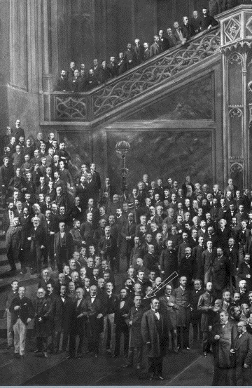
- Une
leçon de Charcot à La
Salpêtrière, tableau de M
Brouillet
- Œuvres
principales de Charcot
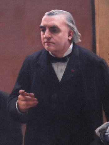
- Charcot
JM The topography of the brain Forum
1886
- Charcot
JM Magnetism and hypnotism Forum
1889
- Hypnotisme
and crime Charcot JM
1890
-
- Les
internes de JM. Charcot
-
- JM
Charcot et une patiente
ataxique
1875 la seule photo connue de
Charcot avec un malade
-
- Croquis
de JM. Charcot par Paul
Richer
-
-
- Charcot
in Morocco: Introduction, notes and translation
by Toby Gelfand
-
|
