Encephalitis
lethargica syndrome: 20 new cases and evidence
of basal ganglia
autoimmunity
RC
Dale, AJ Church, RA H. Surtee, AJ Lees, JE
Adcock,
B Hardin, BG Neville, G
Giovannoni
1 Neurosciences Unit
and 2 Neuropathology Department, Great Ormond
Street Hospital and Institute of Child Health, 3
Department of Neuroinflammation, Institute of
Neurology, University College London, 4 Reta
Lila Weston Institute of Neurological Studies,
Royal Free and UCLMS, London and 5 Department of
Neurology, Radcliffe Infirmary, Oxford,
UK
Case example 2
A 15-year-old boy presented with an acute
personality change 10 days after an upper
respiratory tract infection. He became extremely
anxious and worried about his safety. One week
later he had an oculogyric crisis and developed
upper-limb resting tremor and bradykinesia. This
was followed by extreme daytime somnolence,
lethargy and intractable hiccough. On
examination, he would fall asleep if not
stimulated and yawned continuously.
Pupillary responses were poorly reactive to
light and accommodation. There was tongue
tremor, a positive glabellar tap and slow
speech. Limb examination revealed rigidity with
cogwheeling, bradykinesia and freezing. He had a
stooped gait with poor arm-swing. Positive
results included an elevated ASOT (350 IU/ml)
and a mirrored pattern of OCB in both CSF and
serum. PCR of CSF for neurotropic viruses was
negative. MRI of the brain showed enhancement of
the basal ganglia. He was treated with 50 mg of
levodopa twice a day (with carbidopa), which
improved his sleep disorder and parkinsonian
signs, although he complained of insomnia. His
abnormal clinical signs remained for 2 months,
following which the levodopa was withdrawn. He
has made a complete recovery with no
neurological or psychiatric sequelae at 1 year
of follow-up.
Abstract : In 1916, von
Economo first described encephalitis
lethargica (EL), a CNS disorder presenting
with pharyngitis followed by sleep disorder,
basal ganglia signs (particularly parkinsonism)
and neuropsychiatric sequelae. Since the
1916Ð1927 epidemic, only sporadic cases have
been described.
Pathological studies revealed an
encephalitis of the midbrain and basal ganglia,
with lymphocyte (predominantly plasma cell)
infiltration. The EL epidemic occurred during
the same time period as the 1918 influenza
pandemic, and the two outbreaks have been linked
in the medical literature. However, von Economo
and other contemporary scientists thought that
the 1918 influenza virus was not the cause of
EL. Recent examination of archived EL brain
material has failed to demonstrate influenza
RNA, adding to the evidence that EL was not an
invasive influenza encephalitis.
By contrast, the findings of intrathecal
oligoclonal bands (OCB) and beneficial effects
of steroid treatments have provoked the
hypothesis that EL may be immune-mediated. We
have recently seen 20 patients with a similar EL
phenotype, 55% of whom had a preceding
pharyngitis. The patients had remarkable
similarity to the historical descriptions of EL:
sleep disorder (somnolence, sleep inversion or
insomnia), lethargy, parkinsonism, dyskinesias
and neuropsychiatric symptoms. CSF examination
commonly showed elevated protein and OCB (75 and
69% respectively).
Investigation found no evidence of viral
encephalitis or other recognized causes of
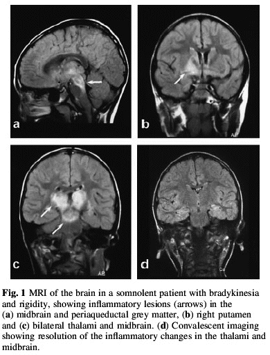
rapid-onset parkinsonism. MRI of the brain was
normal in 60% but showed inflammatory changes
localized to the deep grey matter in 40% of
patients. We investigated the possibility that
this phenotype could be a postinfectious
autoimmune CNS disorder, and therefore similar
to Sydenham's chorea.
Anti-streptolysin-O titres were elevated in
65% of patients. Furthermore, western
immunoblotting showed that 95% of EL patients
had autoantibodies reactive against human basal
ganglia antigens. These antibodies were also
present in the CSF in four patients tested. By
contrast, antibodies reactive against the basal
ganglia were found in only 2Ð4% of child and
adult controls (n = 173, P < 0.0001). Rather
than showing polyspecific binding, these
antibodies bound to common neural autoantigens
of molecular weight 40, 45, 60 and 98 kDa.
Regional tissue comparisons showed that the
majority of these autoantigens were specific to
or enriched in CNS tissue. Immunohistochemistry
with secondary staining localized antibody
binding to neurons rather than glial
populations. Further investigation is required
to determine whether these antibodies affect
neuronal function (i.e. whether they are
pathogenic anti-neuronal antibodies).
Histopathology in one case demonstrated striatal
encephalitis with perivenous B- and
T-lymphocytic infiltration. We believe an
EL-like syndrome is still prevalent, and propose
that this syndrome may be secondary to
autoimmunity against deep grey matter
neurons.
Encephalitis
lethargica: part of a spectrum of
post-streptococcal autoimmune
diseases?
Brain January 2004; 127; 1;
2-3, Editorial
Angela Vincent
Neurosciences Group,
Department of Clinical Neurology, Weatherall
Institute of Molecular Medicine, John Radcliffe
Hospital, Oxford UK
Encephalitis lethargica (EL) was first
described by von Economo in 1917, shortly after
the start of the 1916Ð1927 epidemic. The
patients, mostly children of either sex,
characteristically presented with headache and
malaise, lethargy, insomnia, and
ophthalmoplegia. Some recovered but the others
either died during an acute fulminating disorder
or developed, insidiously or after a variable
period of time, movement and/or psychiatric
disorders including Parkinsonism, oculogyric
crises, chorea, myoclonus, mutism, catatonia or
behavioural problems. Although linked by many
observers to the influenza epidemic, the
epidemic of EL began earlier and lasted longer,
and flu virus has not been found in archival
post-mortem tissue (e.g. McCall et al., 2001; Lo
and Geddes, 2003). Sporadic cases are still
reported, but the acute fulminating form seems
to have disappeared, and there have been no
further reported epidemics.
The paper by Dale and colleagues in this
issue of Brain describes 20 patients with a
condition presenting with sleep disorder,
lethargy, Parkinsonism and neuropsychiatric
disorders, including mutism, anxiety,
depression, obsessions and compulsions (Dale et
al., 2004). The patients ranged between 2 and 69
years of age, but most were children or
teenagers. Although we are not told the time
between onset and study by the authors, half of
the patients had a monophasic illness, and five
have made a good recovery, but the others have
continuing problems of movement or
neuropsychiatric disorders at a follow-up period
of <2 years. The clinical features, the
course of the disease, cerebrospinal fluid and
imaging studies, and histology in one case, are
similar to those in cases of EL as originally or
subsequently described, except that
ophthalmoplegia and oculogyric crises were only
found in a minority of Dale's cases. The 20
cases were referred from tertiary neurological
centres over a period of 3 years, suggesting a
somewhat higher incidence of this EL-like
illness than is apparent from the recent
literature.
The main emphasis in their paper is the
evidence for an autoimmune, possibly
post-streptococal, aetiology. High signal
changes were found on T2 imaging in 40% of the
cases, predominantly in the deep grey matter,
which resolved in a few cases examined during
convalescence. Oligoclonal bands (OCB) were
found in nine of the 13 examined; in five cases
OCB were restricted to the cerebrospinal fluid
(CSF) indicating intrathecal synthesis. These
findings are consistent with those reported in
sporadic cases; for instance, a recent case of
EL showed plasma cell infiltrates in the brain
and very high IgG levels in the CSF (Kiley and
Esiri, 2001). But in four cases the OCB were
detected in both CSF and serum, which suggests
an immune response originating in the periphery.
Indeed about half of the patients had a previous
infection, either of the upper respiratory tract
or tonsillitis, and raised titres of
anti-streptolysin-O antibodies were present in
65%. Streptococcal infections were also present
in some of the original patients with EL, and
similarities to Sydenham's chorea were noted
during the epidemic. Remarkably, an EL like
illness was induced in dogs by vaccination
against streptococcus (von Economo, 1931).
Several authors have previously detected
antibodies to neuronal antigens in neurological
diseases associated, at least in part, with
streptococcal infections including Sydenham's
chorea and PANDAS (paediatric autoimmune
neuropsychiatric disorders associated with
streptococcal infections; Kiessling et al.,
1993; Singer et al., 1998; Church et al., 2002).
Indeed, one recent study suggests that such
antibodies to streptococcal antigens cross-react
with lysogangliosides on the neuronal cell
surface (Kirvan et al., 2003) In the present
study Dale et al. (2004), detected antibodies,
similar to those they have previously described
as anti-basal-ganglia antibodies, by western
blotting of soluble extracts of basal ganglia
homogenates from human brain. The antibodies
were detected at very low frequency in control
groups but were found in 95% of EL sera and 4/5
of the CSFs tested. They bound to several
different polypeptide bands, 40, 45, 60 and 98
kDa, of which the 40, 45 and 60 kDa bands
appeared to be the same as those found
previously, by the same team, in other
post-streptococcal conditions. This raises
issues of disease specificity but would be
consistent with their hypothesis that EL is part
of a spectrum of immune-mediated
post-streptococcal basal ganglia disorders.
Western blotting is often used as a first
test for anti-neuronal antibodies, but although
it efficiently detects antibodies to
non-conformational epitopes, it is likely to
miss those potentially pathogenic antibodies
that bind to conformational determinants.
Moreover, it would have been more informative to
use a whole tissue or membrane preparation,
rather than soluble extract, since antibodies
that exclusively recognise membrane targets
would have been missed in the latter procedure.
To see, therefore, whether there were serum
antibodies binding to intact tissue, they
performed immunofluorescence which demonstrated
binding of antibodies to axons and neuronal cell
cytoplasm. The results indicate the presence of
antibodies predominantly to intracellular
targets but do not exclude the possibility that
there were also antibodies binding to the
neuronal cell membranes. There are, however,
questions regarding the regional specificity of
the antibodies since a careful analysis of
different parts of the brain was not done.
Binding to bands in homogenates of whole rat
brain and rat cerebellum were said to be
similar, suggesting that the term 'anti-basal
ganglia antibody' may prove to be
misleading.
Thus there are some concerns about the
neuronal specificity and pathogenic relevance of
the antibodies detected, as the authors
acknowledge. The presence of antibodies to
different protein bands, varying between the
individual patients, suggests that they may be
secondary to an immune mediated condition rather
than causative. In addition, it is not yet clear
whether the antibodies to the basal ganglia
antigens are cross-reactive with streptococcus A
antigens. One would like to see, in future
studies, evidence for antibodies binding to the
surface of intact neurones derived from rat
basal ganglia preparations, or other brain
regions for comparison, and absorption
experiments with streptococcal antigens to
demonstrate cross-reactivity. Moreover, serial
studies on individual patients comparing
antibodies to streptococcal and neuronal
antigens should be performed.
The two most convincing criteria of an
antibody-mediated disorder, even in the absence
of the detection of specific antibodies, are the
response to immunosuppressive therapies and
passive transfer of disease to experimental
animals. PANDAS has been shown to respond to
plasma exchange (Perlmutter et al., 1999), and
corticosteroids have been found effective in
individual cases of EL (e.g. Blunt et al., 1997)
although they were not apparently tested in
these patients. Recently, a Tourette's-like
syndrome has been transferred to rats by
intracerebral injections (Hallett et al., 2000;
Taylor et al., 2002). If EL is indeed
immune-mediated, it should be possible to show a
clinical response to plasma exchange and other
immunotherapies, at least early in the course of
the disease before permanent changes take place,
and successful transfer of disease to
experimental animals by immunoglobulin
injection. Although passive transfer is still
not routinely performed for CNS disorders, the
growing number of conditions in which an
antibody-mediated aetiology is now suspected
(reviewed in Lang et al., 2003), and preliminary
studies in which functional effects of
antibodies on CNS neurons are being shown (e.g.
DeGiorgio et al., 2001; Kirvan et al., 2003),
means that further approaches to demonstrate the
roles and mechanisms of action of
peripherally-induced antibodies in causing CNS
diseases need to be developed. However, one
should not forget the possibility that the
anti-neuronal antibodies may be markers for a
destructive process, either T cell-mediated or
due to direct toxicity from the infectious
agents or the activated immune system. This
paper, in suggesting a role for the immune
system in EL and in other disorders that appear
to form part of a spectrum of post-streptococcal
autoimmune diseases, highlights the need for
more experimental work in this important
area.
References
Blunt SB, Lane RJ, Turjanski N, Perkin GD.
Clinical features and management of two cases of
encephalitis lethargica. Mov Disord 1997; 12:
354-9
Church AJ, Cardoso F, Dale RC, Lees AJ,
Thompson EJ, Giovannoni G. Anti-basal ganglia
antibodies in acute and persistent Sydenham's
chorea. Neurology 2002; 59: 227-31.
DeGiorgio LA, Konstantinov KN, Lee SC,
Hardin JA, Volpe BT, Diamond B. A subset of
lupus anti-DNA antibodies cross-reacts with the
NR2 glutamate receptor in systemic lupus
erythematosus. Nat Med 2001; 7: 1189-93.
Kiessling LS, Marcotte AC, Culpepper L.
Antineuronal antibodies in movement disorders.
Pediatrics 1993; 92: 39-43.
Kiley M, Esiri MM. A contemporary case of
encephalitis lethargica. Clin Neuropathol 2001;
20: 2-7
Kirvan CA, Swedo SE, Heuser JS, Cunningham
MW. Mimicry and auto antibody-mediated neuronal
cell signaling in Sydenham chorea. Nat Med 2003;
9: 914-20
Lang B, Dale RC, Vincent A. New autoantibody
mediated disorders of the central nervous
system. Curr Opin Neurol 2003; 16: 351-7
Lo KC, Geddes JF, Daniels RS, Oxford JS.
Lack of detection of influenza genes in archived
formalin-fixed, paraffin wax-embedded brain
samples of encephalitis lethargica patients from
1916 to 1920. Virchows Arch 2003; 442:
591-6.
McCall S, Henry JM, Reid AH, Taubenberger
JK. Influenza RNA not detected in archival brain
tissues from acute encephalitis lethargica cases
or in postencephalitic Parkinson cases. J
Neuropathol Exp Neurolol 2001; 60: 696-704
Perlmutter SJ, Leitman SF, Garvey MA,
Hamburger S, Feldman E, Leonard HL, et al.
Therapeutic plasma exchange and intravenous
immunoglobulin for obsessiveÐcompulsive
disorder and tic disorders in childhood. Lancet
1999; 354: 1153-8
Singer HS, Giuliano JD, Hansen BH, Hallett
JJ, Laurino JP, Benson M, et al. Antibodies
against human putamen in children with Tourette
syndrome. Neurology 1998; 50: 1618-24
Taylor JR, Morshed SA, Parveen S, Mercadante
MT, Scahill L, Peterson BS, et al. An animal
model of Tourette's syndrome. Am J Psychiatry
2002; 159: 657-60
von Economo C. Encephalitis lethargica. Its
sequelae and treatment. Translated by K. O.
Newman. London: Oxford University Press;
1931.
Encéphalite léthargique
Cruchet, Moutier, Calmettes Soc méd hop
Paris 27 avril 1917